Some animals have been farming for much longer than humans.
Thousands of years ago, farming revolutionized human societies. It allowed us to settle in one place and to have surplus food, freeing some people to pursue other specialized labor. Populations grew, cities flourished. Today, it is impossible to think about life without agriculture. But did you know that some animals have been farming for much longer than us?
Insects are probably the most well known farming animals, but accomplished farmers have been documented among fish, crustaceans, and mollusks as well. Even microorganisms have been reported to farm. In 2011 a group of researchers reported on the amoeba Dictyostelium discoidelium, which farms bacteria.
Some animals practice very advanced forms of agriculture, which include preparing the growth medium, propagating their crop, tending their gardens, fertilizing and harvesting the crop, and transmitting it from parents to offspring. The most advanced forms of animal farming belong without a doubt to the insects, but many other animals practice simpler farming techniques too.
Meet some of these amazing animals below, and stay tuned to Part 2 of this series for a look into the more advanced farming species.
In 2005, researchers discovered a new species of crab, Kiwa hirsuta, at a hydrothermal vent in the South Pacific Ocean. It was a bizarre creature, covered in hair-like structures called setae, which earned it the common name of Yeti crab. However, only one specimen was collected, and many questions remained about the crabs’ diet and feeding strategy.
Then, in 2006 a second Yeti crab species was discovered off the coast of Costa Rica, on a methane seep. These crabs, Kiwa puravida, displayed an odd habit of rhythmically waving their claws over areas of methane seepage (see video below).
Examination of different specimens revealed that the hairy structures on the crabs’ claws were covered by chemosynthetic epibiotic bacteria. By studying the composition of the crabs’ tissues, researchers were able to determine that these bacteria constitute the Yeti crabs’ main food source. The crabs fertilized their bacterial crops by swaying their claws over the methane seepage sites, allowing nutrients to wash over the bacteria. They then use a specialized mouth appendage to transfer the bacteria from the setae to their mouths.
Video S1: Kiwa puravida at Mound 12, Costa Rica demonstrating the rhythmic waiving of its chelipeds (Thurber et al., 2011).
In 2015, a third species of Yeti crab was discovered living on hydrothermal vents in the Antarctic Ocean, Kiwa tyleri. Dense setae covering its ventral side give it the appearance of a hairy chest, for which it has been nicknamed the “Hoff crab“, after the actor David Hasselhoff.
Damselfishes are a group of small to medium fish living mostly in saltwater ecosystems, and many of which inhabit the world’s tropical coral reef systems. They have a reputation for being feisty and territorial. Damselfishes feed on small crustaceans and other small organisms, sponges, and algae. Some algae- eating damselfish species are actually accomplished gardeners, tending carefully to their algae garden, from which they feed.
The damselfish Sterogastes nigricans. Photograph by Hata et al., BMC Evolutionary Biology.
The damselfish Stegastes nigricans,a little fish with an unassuming appearance, is one such dedicated farmer. It inhabits tropical coral reefs between 30°S and 30°N of the equator, and assiduously keep patches of the filamentous red algae Polysiphonia sp. It keeps watch over his algae patch aggressively, weeding out other algae species, and defending the patch against other herbivores. Researchers observed that when damselfishes were removed, other species of algae readily invaded the Polysiphonia patches.
Although this behavior is beneficial for both damselfish and algae, there might be some negative effects for the health of the coral reef. In its effort to create space for its algae garden, the aggressive weeding by the damselfish prevents coral polyps from attaching. Also, the algae in their garden can harbor bacteria associated with coral disease. In a 2014 research paper, Casey et al. reported greater bacterial concentration and incidence of black band disease within S. nigricans territories than outside.
Littoraria irrorata, also known as the Marsh periwinkle, is a species of salt-water snail that lives in salt marshes on the Atlantic and Gulf coasts of North America. This surprising little farmer grazes on stalks of cordgrass, but not to eat. Rasping with its radula, it creates longitudinal grooves on the stalks. Opening up the plant’s tissues facilitates invasion by ascomycete fungi, which are abundant in the salt marsh environment, and are the periwinkle’s preferred food.
These snails have even been observed fertilizing their fungus crops with their feces, which are rich in nutrients and fungus hyphae.
Source:
Silliman, B.R. and Newell, Y. 2003. Fungal farming in a snail. Proceedings of the National Academy of Sciences, 100(26): 15643-15648.
These are just a few examples of animals that have developed farming relationships with their food, a type of symbiosis referred to as cultivation mutualism. In the next post, we’ll learn about some of the more advanced animal farming societies.
Check out some of last week’s most interesting news stories:
1. Generating Power from Polluted Air
This small device can generate hydrogen fuel while purifying polluted air. Photograph by UAntwerpen and KU Leuven.
Air pollution is a major environmental issue, and a cause of health problems affecting millions of people globally. Fuel combustion is one of the main causes of air pollution, and scientists are earnestly trying to find solutions for air remediation, as well as alternative energy sources that are more environmentally friendly. Researchers from Belgium recently reported on a way to do both using a single device, which can turn air pollution into fuel. And it gets even better, it needs only light to operate.
It is a very simple device, consisting of two chambers separated by a membrane. The membrane is made of special nanomaterials that act as catalysts. Contaminants are degraded on one side, and hydrogen gas is produced on the other. The gas is stored and can be used for fuel. The device is currently very small, but the researchers are working on scaling up the technology.
Verbruggen, S.W., et al. ChemSusChem. DOI: 10.1002/cssc.201700485.
2. Spray-Painting Touchpads
Researchers at Carnegie Mellon have shown that touchpads need not be restricted to the flat surfaces of phones and tablets anymore.
No longer will touchpads be restricted to flat phone or computer screens. Researchers at Carnegie Mellon have figured out a way to turn almost any kind of surface into a touchpad. The technology, which they presented at the Conference on Human Factors in Computing Systems last week, is based on the use of an electrically conductive coating that can be applied as easily as spray painting. When a finger touches the surface, some of the electric current is steered to ground through the hand, temporarily lowering the voltage. The researchers showed that it is possible to localize, with up to 1 cm of precision, where and when this occurs by using electrodes connected around the edges of the coating.
Apart from spray painting, other methods like 3D printing can be used to apply the coating, or even make the entire object using the conductive material. The researchers have already used the technology to create touchpads on several different surfaces, including a steering wheel, a guitar, and even jello. The possibilities are endless. Check out the video below to see how it works.
Video by Future Interfaces Group, Carnegie Mellon University.
Recently, researchers at the University of Minnesota developed a 3D printed flexible electronic sensory device that could be used to give robots the ability to sense their environment. It consists of several layers, including a bottom silicone surface, and top and bottom electrode layers separated by a pressure sensor shaped like a coil.
An interesting application of these sensory devices would be to put them on surgical robots, which would allow the surgeon to feel during the operation. Furthermore, because the material can be printed at low temperatures, it could be used to print on skin. This opens up many practical uses for these sensors, such as printing wearable patches for health monitoring.
Visit the University of Minnesota webpage for more details, as well as a video of the printer in action.
Researchers at Lancaster University are making it easier to turn used coffee grounds into fuel.
Did you know that used coffee grounds can be used to produce biodiesel? Biofuels produced from feedstocks are generally not the ideal replacement for fossil fuels, because of all the resources required to grow them. Used coffee grounds, however, offer a convenient, low-cost alternative. Unfortunately, their commercial competitiveness is negatively affected by high processing times and costs. Consequently, very few companies are currently taking advantage of this potential biofuel source, and most of the spent coffee grounds that are produced daily end up in landfills.
In a study published earlier this month, scientists from Lancaster University figured out a way to make the process much more efficient than what was previously used. While the technique typically involves two steps, one for extracting the oils and another for turning them into biodiesel, they showed that it is possible to do both steps at once. This modified process dramatically cuts down on processing times and costs, and will hopefully lead to a greater exploitation of this alternative fuel source.
Cricket farm in Thailand. Photograph by Afton Halloran.
It is well known that meat production places a high burden on the environment. Insects have often been called the food of the future because of their high nutritional value, and potentially lower environmental impact. Recently, a study on the sustainability of crickets as a food source showed that they can in fact be produced in a more environmentally friendly way than other livestock. The study compared the environmental impact of cricket and broiler chicken production in Thailand, by focusing on certain ecological markers, such as eutrophication and resource consumption.
They found that in most of the markers studied, cricket farming had a smaller ecological footprint. In both cricket and chicken farms, feed production was one of the areas with the highest impacts. Although it is not their natural diet, farmed crickets are fed chicken feed because it makes them grow faster. However, crickets are more efficient in turning the feed into animal protein, thus resulting in a lower impact than chickens. Researchers are now working on finding better feed sources for commercially farmed crickets in order to reduce the ecological footprint of raising them even more.
To learn more about the story, visit the University of Copenhagen website.
Check out this week’s summary of some of the most interesting science news:
1. Killing Bacteria with…Paper?
Flexible, paper-based plasma generators can inactivate microbes within seconds (Photograph by Jingjin Xie).
Scientists from Rutger’s University have developed a new flexible, portable and disposable device for killing bacteria using metallized paper. It consists of stacked layers of paper covered with aluminum in a honeycomb pattern. When high voltage is applied, plasma is generated. Plasma is a mixture of ions, reactive molecules and UV radiation, which is regularly used to kill bacteria. In these devices, the stacked, porous paper allows gas to permeate it, which in turn is fuel for the plasma and allows for cooling. The researchers showed they were able to inactivate 99.9% of E. coli cells after just 30 seconds of treatment. Further research is needed to assess how effective these devices are at eliminating spores, but preliminary results look promising.
In the future, this specialized paper could be used to clean laboratory equipment, as sanitizing bandages for wounds or as protective layers in clothing. Part of the researchers’ motivation was to develop clothes that could self-sterilize after being exposed to pathogens, like in the case of health workers during the ebola epidemic of 2014.
Researchers from Stanford University developed a new flexible, biodegradable semiconductor, shown here on a human hair (Photograph from Bao Lab).
With the short lifespan and accessibility of electronics, electronic waste, or e-waste, is quickly becoming an ecological problem. Electronic circuits are made from non-biodegradable materials, often containing toxic substances like heavy metals. Looking for potential solutions to this mounting problem, a group of researchers from Stanford University developed a flexible, biodegradable semiconductor polymer that decomposes when exposed to mild acids. In addition, they developed a biodegradable electronic circuit, and a degradable substrate to hold everything together. The film-thin substrate is made from cellulose and can stick to rough and smooth surfaces. The best part is, when the circuit is no longer needed, everything can easily be degraded.
This new electronic device has the potential to be used in a variety of applications, including wearable electronics, like skin sensors to monitor glucose or blood pressure, and even implantable devices.
3. Water-Repellent Material Inspired by Snake Skin
In yet another example of scientists being inspired by nature, a research group from the University of Freiburg, in Germany, has developed a new water-repellent material that, when damaged, is capable of shedding its outer layer, much like the skin of a snake. The material comprises three layers, an upper water-repellent material, a middle layer made from a water-soluble polymer, and a super-hydrophobic silicon layer at the bottom. When the top layer is scratched and the material is submerged in water, the middle layer is dissolved and the top layer peels back, exposing the hydrophobic layer underneath.
Current water-repellent materials are sensitive to mechanical damage, leading to water permeation. In contrast, this new development could lead to the development of self-renewing water-repellent materials, like cloth for raincoats, that could restore their own hydrophobicity after damage. Check out the video below to see this snakeskin-like material in action.
4. “Princess Pheromone” Is Expressed by Future Queen Ants
Adult working ants carefully inspect larvae to detect which show signs of becoming queens (Photograph by Clint Penick)
Every summer, around the time of the first rains, new queens of Indian jumping ants, Harpegnathos saltator, develop. Worker ants carefully groom them until they are ready to mate with the flying males, and later they leave to found their own colonies. But how do the workers distinguish between would-be queens and normal worker larvae? In a new study, led by Clint Penick from North Carolina State University, researchers identified a pheromone, which they call “princess pheromone,” produced by larvae that are preparing to become queens. This chemical communication allows adult ants to know which larvae are turning into queens, and thus work to ensure their proper development.
Interestingly, when the princess pheromone is expressed at inappropriate times, adult ants respond in a radically different fashion. When the timing is off for mating, or when there are already too many queens in development, adults respond by biting the larva expressing the pheromone. Harassed into submission, the larva subsequently develops into worker. This behavior allows for conservation of resources, as well as preservation of colony structure by preventing the development of more queens than the colony can support.
High-resolution satellite imagery offers a less disruptive and more cost-efficient alternative to keep track of albatross colonies in remote islands.
The albatrosses are a group of large, marine birds comprising several species, a number of which are critically endangered. It is difficult to keep track of their population numbers, because they nest in remote islands that are often unpopulated and hard to reach. But now, in a study published in the journal Ibis, scientists show that the highest resolution satellite images can capture individual albatrosses, making it possible to count them without actually having to be there.
In this study, they used 30-cm resolution images from the WorldView-3 satellite to count a Wandering Albatross colony in South Georgia, a well-studied group, and found the numbers comparable to ground-counts of nests. They then applied the technique to count a remote colony of endangered Northern Royal Albatross in the Chatham Islands, which had not been counted recently, and made a concerning discovery. On one of the islands, the satellite count was significantly lower than the previous ground-count, which illustrates the importance of frequent population counts for conservation efforts. The use of this new satellite imagery offers a great alternative for better and less disruptive monitoring of bird populations, which is especially important for endangered species.
An ecosystem is a complex web of interactions between different organisms and their environment. The interactions between species have often been tuned by years of co-evolution, resulting in specialized defense mechanisms and adaptations to their specific environment. When a non-native species is introduced, it has the potential to become very disruptive.
How are exotic species introduced?
The introduction of non-native species is often an unwanted result of human activity. Ever since humans started exploring and colonizing new lands, hitchhiking organisms have accompanied them. Perhaps the most famous example of this has been the spread of the common house rat Rattus rattus, native to Asia and now a worldwide pest. Alien species are still spread today through human transportation systems, like when fish and marine invertebrates are carried in the ballast water of ships.
Other times, nonnative species are deliberately introduced. For example, rabbits were released in Australia in the 1800s for sport and have since become widespread, to the detriment of Australian ecology. In other cases, animals are liberated into the wild by well-meaning pet owners who no longer wish for or are able to care for their exotic pets.
To be sure, not every alien species turns into a threat, or even manages to survive. Introduced organisms have not adapted to cope with the new site’s climate, predators and competitors. Moreover, if they were previously kept in captivity they might be ill-suited to survive on their own, and are likely to die from starvation, depredation or exposure. However, some species manage to survive and even thrive, often at the expense of native organisms. Whether an alien species becomes invasive or not depends on several factors, including lack of natural predators or diseases, flexibility to adapt to different environmental conditions, dispersion and reproduction rates, and diet diversity.
An invasive species, then, is any non-indigenous animal, plant, fungus, or bacteria, that becomes established in a new environment, and threatens or damages resident species. In fact, invasive species are considered one of the top causes for loss of biodiversity worldwide, and can lead to the extinction of native species.
Why are invasive species harmful?
Invasive species may outcompete native organisms for food or shelter. Native species often don’t have defense mechanisms against introduced species, and so can easily become prey. Invasive species can also modify the environment in a way that is detrimental for indigenous organisms. For example, the common carp, native to Eurasia but introduced in America, increases the turbidity of water and uproots vegetation due to its eating habits, affecting native fish that need clear water to survive. Other times, invasive species may harbor pathogens for which the native organisms have no adequate defenses. A concerning example of this is the case of the European salamanders, which are rapidly disappearing as a result of the fungus Batrachochytrium salamandrivorans, likely introduced through the pet trade.
Read on to learn about some of the most important invasive species affecting our ecosystems today:
1. Lionfish
Two invasive species of lionfish are threatening the health of Caribbean coral reefs.
Lionfish are fish with showy fins and venomous spikes, popular among aquarists. They are native to the Indo-Pacific Ocean, but two species, Pterois volitans and Pterois miles, have become very successful invasive species in the Eastern Atlantic Ocean and the Caribbean Sea. They were first observed off the coast of Florida in the 1980s, and have since spread along the East coast of the US and throughout the Caribbean, with some specimens found as far south as Brazil. There have even been some sightings as far north as Rhode Island, although they are not suited for cold temperatures. It is generally accepted that the lionfish invasion was a result of several introductions, including specimens released by aquarists. Several lionfish were also reportedly released into the wild from a flooded aquarium after Hurricane Andrew in 1992.
There are several reasons why the lionfish invasion has been so successful. They are aggressive fish with a voracious appetite and a very diverse diet. They are also able to spread widely using ocean currents for the dispersion of their larvae. Lionfish have few natural predators thanks to the protection of their venomous spikes. As a result, they have become an important threat to the health of the native coral-reefs and fish communities. In a study on lionfish eating habits off of the Bahamas, scientists found that during the period from 2004 to 2010, lionfish numbers increased considerably, to the point of constituting 40% of predator biomass in the area. Meanwhile the biomass of the 42 species of fish identified as prey for the lionfish decreased by 65%. Current control efforts include spearfishing, and promoting lionfish meat as a delicacy.
2. Cane Toad
Cane toads are an invasive species in more than 30 countries.
The cane toad, Rhinella marina, is a species of toad native to Central and South America. It is a large toad with a blotched, warty skin that produces a toxic secretion. In the early 20th century, cane toads were introduced in Australia and several islands of the Caribbean and Oceania as biological control for pests plaguing sugarcane plantations. However, the toads soon became pests themselves.
Cane toads breed prolifically and have a long lifespan, living up to 15 years in the wild. They have a voracious appetite and a broad diet, which includes rodents, reptiles and other amphibians, birds, invertebrates, plants, dog food, and even hosehold waste. In addition, they have a powerful protection against predators. Adult toads have glands that release potent toxins, and tadpoles are toxic if ingested. The toads are detrimental to many native species of reptiles, with which they compete for food, as well as for frog-eating predators, which can become intoxicated.
3. American Mink
The American mink is an invasive species in Scotland, where it threatens native bird populations.
American mink, Neovison vison, were brought into Scotland in the 1950s to be farmed for their fur. It is not clear whether some individuals escaped the farms, or if they were intentionally released, but the minks found their way out into Scottish wilderness and proliferated, causing great damage to native species. The American mink is a very adept hunter, and eats fish, small mammals, birds, and amphibians. Particularly vulnerable are species of ground-nesting birds such as moorhens and arctic tern, and small mammals like the water vole. The American mink also causes significant losses for farmers by taking domestic chickens and ducks. Fortunately, aggressive campaigns to eradicate this furry fiend from key locations have been put in place successfully, and many of the local species are making a comeback.
4. Burmese Python
The Burmese python is an invasive species in Florida.
The Burmese python, Python bivittatus, is one of the large constrictor snake species, measuring on average 3.7 m. It is often found on land near bodies of water, but can also climb trees, especially as a juvenile. Their native range is South and Southeast Asia, but they are an invasive species in the Everglades National Park, in South Florida. The introduction of pythons is thought to have occurred when specimens escaped python farms as a result of Hurricane Andrew in 1992, as well as through the release of python pets.
In the Everglades, they hunt and eat birds, mammals, and alligators. Their diet is varied and has been known to include endangered native species. In fact, the spread of pythons in the Everglades has been associated with the decline in several species of mammals, including raccoons, opossums, bobcats and rabbits. Efforts to control the python population include annual capturing competitions, and the use of trained dogs to spot and capture the snakes.
5. Walking Catfish
Walking catfish is the common name of Clarias batrachus, a species of freshwater catfish native to Southeast Asia. It is an air-breathing fish, and gets its name from its ability to “walk” on its pectoral fins while wiggling its tail (see video below). The fish uses this ability to look for food and new bodies of water. It was introduced in Florida in the 1960s, and has since become an invasive species.
The walking catfish has a voracious appetite and a diverse diet, which includes other fish, invertebrates and plants. They sometimes enter aquaculture farms, going from pool to pool preying on the captive fish. They are also known to harbor the bacteria for enteric septicemia, which can infect farmed fish. These catfish are able to thrive even in small, stagnant pools that are often low in oxygen. They become most aggressive in small pools during the dry season, and can easily become dominant. During cold months or periods of drought, they burrow in the mud and become dormant, emerging when the weather turns more favorable.
6. Water Hyacinth
The water hyacinth can quickly crowd bodies of water when not controlled.
The water hyacinth, Eichhornia crassipes, is an aquatic plant native to the Amazon. It is characterized by floating leaves and pretty flowers that range in color from lavender to pink. The water hyacinth reproduces very rapidly. New plants can be formed by runners from parent plants, or through seeds. Because of their prolific growth, the water hyacinth tends to cover entire bodies of water when not controlled. This is a problem, because it blocks sunlight from reaching other native plants, and removes oxygen from the water, killing the fish. Furthermore, they create a favorable environment for mosquito and snail reproduction, which are known vectors for diseases, including malaria. Water hyacinth populations are known to double their biomass in as little as 12 days. Their uncontrolled growth also clogs waterways and reduces access for boats, fishing, and other human activities.
Their pretty flowers make them popular ornamental plants, which is probably the pathway that has lead to the introduction of this species in foreign ecosystems. The seeds of the water hyacinth can survive for a long time which, along with its rapid growth rate, makes it very difficult to completely exterminate this plant. The water hyacinth is a pernicious species in Lake Victoria, the largest African lake and one of the largest lakes worldwide. Eradication efforts began in the 1990s, but a recent resurgence is causing important economic losses, making fishing and water access difficult.
What can we do to help?
The Nature Conservancy lists some simple actions to help combat and prevent the spread of invasive species:
Avoid keeping exotic plant species in your garden
Report sightings of non-native species or, even better, volunteer to help remove invasive species in your area.
Never release unwanted pets into the wild. Not only is it inhumane for the pet, it can be devastating for the ecosystem. Do extensive research if you plan on keeping exotic pets.
Avoid transporting hitchhikers. Check your boots and clothes after a hike to avoid transporting invasive weeds, seeds, or parasites. Check your boat thoroughly before moving it to a different body of water.
It’s one of those scientific discoveries that happened entirely by accident. Federica Bertocchini was busy removing waxworms from her honey beehives when she noticed that the plastic bag where she was collecting the caterpillars had become riddled with holes. The caterpillars were the larvae of the moth Galleria mellonella. These moths lay their eggs in beehives, and when they hatch, the caterpillars feed on the wax.
Back at the lab, Bertocchini and her team confirmed that these caterpillars can indeed digest polyethylene, the most common plastic. Their ability to digest polyethylene could be related to their consumption of wax, since the chemical structure of both compounds is similar. Although polyethylene is a very useful material—its main application is in packaging, including shopping bags—it is creating important ecologic problems. Plastic bags are known to clog landfills and become troublesome in the ecosystems that they enter. Animals often eat them by mistake, or become entangled in them and die. Although these waxworms are not the first organisms discovered to degrade plastic, the rate at which they do it is very surprising. Experiments with plastic shopping bags revealed that 100 worms could consume 92 mg of plastic in 12 hours, which is a rate more than 1000 times higher than that of other previously reported plastic-consuming organisms.
Our gut bacteria might play a bigger role in our lives than we realize.
Several recent studies have linked the bacteria present in the body, the microbiome, with changes in the brain and behavior. Now a group of researchers has found evidence suggesting that gut bacteria can influence animal appetite. Scientists working with the fruit fly, Drosophila melanogaster, recently observed how the presence of certain bacteria species in their gut affects their food preference. When fruit flies are fed a diet deficient in essential amino acids, their reproduction rate markedly decreases and their preference for protein-rich yeast increases significantly. The mechanism through which an amino acid deficiency leads the flies to change their food choices is not entirely understood. In this study, scientists found that bacteria commonly found in the gut of wild fruit flies managed to override the flies’ need to consume protein when fed food that lacked essential amino acids. Even more, the flies that were colonized by these bacteria also didn’t experience a decline in reproduction rate. Surprisingly, the researchers didn’t find evidence that the bacteria provided the lacking essential amino acids to the flies. Furthermore, the flies tended to eat more from food that contained the bacteria than food that didn’t, suggesting that they can alter their feeding patterns to obtain the protective action of the bacteria.
Nature has always been an important source of inspiration for researchers and innovators. This time, scientists developed a new adhesive inspired by the feet of geckos. Geckos, a diverse group of lizards native to warmer climates, are remarkable for their ability to climb up vertical walls and even across ceilings. They can do this thanks to their adhesive toe pads, which are covered in setae, very fine hair-like structures. Different adhesives have been developed mimicking the gecko’s feet, but working on wet conditions has remained a challenge. A potential solution to this problem is the use of hydrogels, which change their stickiness properties by swelling or shrinking depending on the pH. Now, a group of researchers has developed a double-sided adhesive that consists of a membrane covered by hydrogel on both sides. Their experiments showed that the material was capable of switching from high friction and adhesion at a low pH, to low friction and adhesion at high pH, even when wet. A synthetic adhesive capable of working in wet environments opens up a whole range of underwater applications, including underwater robotics and sensors.
4. Baby Humpback Whales Whisper to Avoid Predators
Humpback whale mother and calf
For newborn baby whales, the ocean can be a dangerous place. Humpback whales migrate from their feeding grounds near the north and south poles to warmer tropical waters every year to breed and mate. Afterwards, they return to their higher latitude range with their calves in tow. For a newborn calf, the journey is long and fraught with perils. Exmouth Gulf in Western Australia is a known spot where humpback whale cows and calves rest on their journey south. The researchers tagged mothers and calves to study their behavior without disrupting them. They found that whale calves communicated with their mothers using low volume sounds, much lower than adult humpback whale vocalizations. These sounds, the equivalent of whale whispers, probably to avoid drawing unwanted attention both from male whales who would turn the mother’s attention away from the calf, and from eavesdropping predators. Killer whales are known to hunt humpback calves in the area. Unfortunately, their low-level communications could mean that they are more easily disturbed by encroaching human activities, such as the use of powerboats.
Scientists are working on guidelines to keep astronauts healthy during long missions.
Long-term outer space travel has always been an exciting topic, and now it seems more possible than ever, with plans of manned missions to Mars and even colonizing the moon. In anticipation of this, scientists are working on the publication of a set of medical guidelines for astronauts and all those traveling outside of our planet for extended periods of time.
Researchers from the University of Plymouth and Northumbria University, the Aerospace Medical Association (AsMA), the European Space Agency (ESA), the Royal Air Force (RAF), the International Space University, and Blue Abyss are putting together a Review Group to be launched in May of this year that will systematically analyze all the publications in aerospace medicine. They aim to understand the challenges that astronauts’ bodies will face, such as the effects of microgravity, and will also advice on how to deal with problems the astronauts might face when coming back to Earth. Their conclusions will result in a rulebook intended to help keep all those travelling in space in good health during long trips.
“The enemy of my enemy is my friend,” reads an ancient proverb, and in the fight against antibiotic-resistant bacteria affecting human lives, it is proving true. Earlier this month, researchers from Baylor College of Medicine in Houston published a paper in the journal Scientific Reports, in which they described the use of bacteriophages to combat antibiotic-resistant bacteria. Read on to find out more about what phages are, and why we need them.
The Rise of the Superbugs
Resistance to antibiotics has existed since the development and widespread use of the first antibiotic, penicillin, in the 1940s. In fact Sir Alexander Fleming, the discoverer of penicillin, warned in his Nobel Lecture against the development of resistance that could result from easy access and subsequent incorrect usage of the antibiotic.
Not even the best weapons in our arsenal of drugs can kill some of the superbugs.
As resistance to penicillin inevitably spread, new generations of antibiotics were developed. Bacterial resistance eventually became problematic again, and in the decades that followed, many new antibiotics emerged. However, bacteria are becoming resistant to those as well. We call them “superbugs”, because not even the best weapons in our arsenal of drugs can kill them. Together with a lack of new antibiotic development, this has plunged us into what people have begun calling the “post-antibiotic era,” in which common injuries and infections can once again be life-threatening.
Examples of these superbugs include methicillin-resistant Staphylococcus aureus (MRSA), multi-drug-resistant Mycobacterium tuberculosis (MDR-TB), and carbapanem-resistant Enterobacteriaceae (CRE), like E. coli and Salmonella. CREs, in particular, are resistant to all or nearly all antibiotics available, and around 50% of hospital patients who suffer a blood infection caused by CREs die.
A dramatic example of a superbug infection was reported earlier this year. An elderly woman died of an infection caused by a CRE. She had been in a long-term trip to India, where she broke her femur some years back. She later developed a bone infection, and was hospitalized in India several times. After returning to the US, the woman sought medical care. She was hospitalized in Reno, where it was discovered that she had a CRE infection, in this case by Klebsiella pneumoniae. The bacteria was resistant to 26 different antibiotics, all of the ones that are available in the US. The woman went into septic shock and died shortly after. Even though infections like this one are not common, it is certainly an alarming reminder of what is already starting to happen.
Deaths attributable to antimicrobial resistance (from The Review on Antimicrobial Resistance) / CC BY 4.0
According to the Centers for Disease Control and Prevention, every year at least 2 million people in the United States are infected with antibiotic-resistant bacteria, and at least 23,000 people die from those infections every year. In The Review on Antimicrobial Resistance published in 2014, it is estimated that by 2050 the number of human deaths due to antibiotic-resistant bacteria will be 10 million.
How does resistance develop?
Some bacteria can be naturally resistant to antibiotics. Others may become resistant as a result of random mutations to their DNA. These bacteria then pass their resistance to their offspring. But, even more critically, they can pass their resistance genes horizontally to other bacteria through the exchange of plasmids, or mobile DNA elements. The horizontal spread of resistance can also happen from one species of bacteria to another.
When a bacterial population is exposed to an antibiotic, the susceptible bacteria die. However, if some of them have developed or acquired resistance, they will survive. The continued presence of the antibiotic acts as a selection pressure, killing all susceptible bacteria and allowing the resistant ones to thrive. Overusing antibiotics, as well as using them at a sublethal dose, promotes the development of resistance.
Factors contributing to the crisis
Overprescription of antibiotics, and the widespread lack of regulations regarding their use lead to excessive or unjustified human consumption. As we have seen, such abuse of antibiotics is an important cause of resistance development, but perhaps an even bigger threat is their agricultural use.
Antibiotics are often used in animal husbandry to promote quick growth and prevent infections in crowded conditions.
Although the practice has become increasingly controversial, antibiotics are still commonly used on farm animals, like pigs and chickens, to keep them healthy in crowded environments. They are also used to help the animals grow faster while consuming less food. This allows farmers to increase their yield substantially, but the great amount of antibiotics used, in total greater than that for human consumption, also means these farms are a prime source of resistant bacteria.
Antibiotic-resistant bacteria are present in the feces of these animals, and can become airborne and contaminate the environment. The presence of antibiotic-resistant bacteria has been confirmed in the gut of farm workers, and is also present in slaughterhouses, where the meat of these animals is processed for human consumption. This means that the bacteria can also reach consumers through contaminated meat. Among the resistant bacteria that have been linked to farm animals is Staphylococcus aureus (MRSA).
Adding to this, pharmaceutical companies are less inclined to develop new antibiotics than before. Developing these drugs has become a very expensive and lengthy process for companies, who must comply with very meticulous regulatory standards before being able to market a new drug. The speed with which resistance is developed in bacteria means that some antibiotics are soon obsolete, and many companies have turned their resources to the production of more profitable drugs.
Measures to restrict the use of antibiotics, both for human and animal use, have been established in some places. But as resistance continues to develop faster than new drugs can be created, scientists are beginning to look beyond antibiotics for potential solutions to this looming health crisis.
Bacterio…what?
Bacteriophages are viruses that infect bacterial cells and can destroy whole bacteria populations. They are widespread in nature wherever bacteria are present in high concentrations, like in the intestines of animals and sewages.
The healing properties of the Ganges River first led scientists to investigate the activity of bacteriophages.
The first reports of bacteriophage activity date from the end of the 19th century. In 1896, Ernest Hankin wrote about the healing properties of the Ganges River in India, particularly against cholera. He attributed this property to an unidentified agent, which was small enough to pass through filters and could be inactivated by heat. Later on, in 1915, Frederick Twort described similar findings and suggested several agents that could be responsible, including a virus. However, Twort did not further pursue this line of research, and it was not until a few years later that Félix d’Hérelle, a French-Canadian microbiologist, began to study them seriously. He established that these agents were in fact viruses that could infect and kill bacteria. He named them “bacteriophages,” which means “bacteria eaters.” D’H’erelle also introduced the term page-therapy, or the treatment of bacterial infections with the use of bacteriophages.
Soon, phages started being used on patients, and were even commercially produced in Paris by what is now L’Oreal, and in the US by Eli Lilly. However, the studies analyzing the efficacy of phage therapy were controversial, and this, together with the discovery of antibiotics, led to a decline in interest in Western countries. Phages continued to be used in Eastern countries, particularly in Georgia and Poland, but most of the research was published in non-English languages and was largely unavailable to Western scientists.
Typical structure of a bacteriophage
With the rise superbugs now considered one of the greatest threats to public health, and the lack of new antibiotics, interest in bacteriophages is again rising in the West. Bacteriophages have several advantages over antibiotics. Since superbugs and phages are both present in the environment, phages can evolve and adapt to their hosts in real-time, something antibiotics can’t do. They are also very specific for the type of bacteria they infect, which means that they won’t attack other beneficial bacteria, like our gut bacteria, the way antibiotics do. Furthermore, phages replicate inside their bacterial hosts, which means their dosage will increase inside the body as necessary.
The Research Findings
In the study published by the team from Baylor College of Medicine, the researchers sought to identify a phage for the extraintestinal pathogenic E. coli ST131 (ExPEC ST131). These bacteria are known to colonize the intestinal tract of people without producing symptoms, but can branch out and infect other organs, eventually leading to sepsis and even death. Strains of the type ST131 are resistant to multiple drugs, and are also very virulent. These bacteria are one of the main causes behind antibiotic-resistant E. coli infections in the United States.
The phages were obtained from goose, duck, and canine feces collected from local parks, and chicken feces from a farm. The researchers isolated the phages and tested them against several ExPEC ST131 strains taken from patients. Although no single phage was active against all bacterial strains, the combination of all the phages was.
The researchers then selected one of the promising phages and carried out experiments in mice to evaluate its performance in vivo. After injecting the mice with a virulent strain of ExPEC ST131, followed by a dose of the bacteriophage, they observed that phage-treated mice appeared healthier and had significantly lower bacterial counts than in those that didn’t receive phages.
Human patients with a compromised immune system, such as can be developed as a side effect of chemotherapy, are among the most susceptible to infection by superbugs. Antibiotics are routinely used on these patients, which in addition to promoting bacterial resistance, also eliminate useful gut bacteria , and are not useful with drug-resistant strains.
In this research paper, the this scenario was recreated by allowing a strain of ExPEC ST131 to colonize the intestines of two groups of mice. The researchers then gave the mice alternating daily doses of chemotherapy or phage for six days. Control mice received a placebo instead of the phages. After six days, they counted the bacterial load in several organs and found that it was significantly lower in all organs of phage-treated mice compared to those without phage therapy. The phages were able to lower the bacterial load to levels below what is considered fatal.
Conclusions
As the authors note, the most noteworthy aspect of this study is that they were able to demonstrate that phages against clinically-relevant strains of superbugs can be easily obtained from the environment, and applied in a short time. The fact that at least one of the phages obtained in this way was effective in a mouse model of bacterial infection with a compromised immune system was also encouraging.
Although no single phage was able to kill all the bacterial strains used, the combination of phages from all the sources resulted in killing all the strains. Therefore a cocktail of phages could be relevant in the clinic. The authors also propose the use of phages in combination with antibiotics. Although bacteria can develop resistance to phages, this might cause them to lose their resistance to antibiotics.
Phage therapy, although promising, has some challenges. As mentioned above, it is possible for bacteria to develop resistance to the phages, although phages can also change to adapt to their hosts. However, it has also been observed that the human immune system sometimes develops antibodies to fight the phages, making the therapy ineffective. Notwithstanding, phage therapy certainly seems like an interesting and so-far viable new weapon in our line of defense against bacterial pathogens.
Useful resources to learn more:
Loc-Carrillo, C. and Abendon, S.T. 2011. Pros and cons of phage therapy. Bacteriophage, 1(2), 111-114.
Review on Antimicrobial Resistance. Antimicrobial Resistance: Tackling a Crisis for the Health and Wealth of Nations. 2014.
Sulakvelidze, A., et al. 2001. Bacteriophage Therapy. Antimicrobial Agents and Chemotherapy, 45(3), 649-659.
Ventola, C.L. 2015. The Antibiotic Resistance Crisis. Pharmacy and therapeutics, 40(4), 277-283.
Hi! Here is a brief roundup of some of the week’s most interesting science news. Enjoy!
1. Giant Worm-like Organism Discovered
Giant shipworm Kuphus polythalamia (Credit: University of Utah)
A giant, worm-like mollusk has been described for the first time, and it is the weirdest thing you have seen. The organism, Kuphus polythalamia, is a member of the shipworm family, long bivalved marine mollusks that burrow in wood. It forms a calcareous shell as it burrows into marine sediment. These shells had been observed for years, but the live organism had never been fully described before. The specimens were found in the Philippines, in a muddy marine lagoon full of wood debris. They can grow up to 1.5 meters long, and reach a diameter of 6 cm.
Shipworms normally ingest wood, digesting the cellulose with the help of bacterial symbionts. However, Kuphuspolythalamiarelies on an inorganic chemical for food. It bores into mud, which it sifts through its gills. Bacteria in the gills transform hydrogen sulfide into carbon compounds that the shipworm uses for survival. In fact, the digestive tract of this worm is much shrunken from disuse. The researchers are now studying how the transition from wood-consuming shipworms to giant shipworms that thrive on noxious gas happened.
Researchers at the Fred Hutchinson Cancer Research Center have developed nanoparticles to be used in the fight against cancer. These tiny particles, which are made of a biodegradable material and carry DNA, can penetrate white blood cells and program them to fight cancer cells. A cell-based therapy that has shown promising results in clinical trials involves removing T-cells from the body and manipulating them in the laboratory before implanting them back into the patient. In contrast, the nanoparticles are injected into the patient and used to program the cells inside the body. The DNA-carrying particles attach to the T cells, which ingest them, and deliver their DNA into the cell nucleus. It is like inserting a set of instructions into the cell, which direct it to produce receptors that can identify leukemia cells. The use of these nanoparticles improved animal survival when used in a mouse model for leukemia, from a median survival of 2 weeks to an average survival of 58 days.
Scientists are continually finding potential drugs in the unlikeliest of places. This time, a group of researchers looking into frog slime for compounds exhibiting antimicrobial properties discovered a molecule with anti-flu virus activity. The compound, a peptide they called “urumin” was found in the mucus of the frog Hydrophylax bahuvistara, a species native to Kerala, India. It probably works by binding to hemagluttinin, a protein on the surface of many strains of virus, that the virus needs to in order to infect human cells. Antimicrobial compounds are found in the secretions of many animals, as a form of defense. Frogs are a particularly interesting source because it is relatively easy to obtain the mucus that contains these compounds.
In the fight against antibiotic-resistant bacteria that seem to be becoming more common than ever, scientists are looking beyond antibiotics to combat them. Earlier this week, researchers from the Baylor College of Medicine described in Scientific Reports, how they used bacteriophages, or bacteria-killing viruses, to combat antibiotic-resistant bacteria in mice. They isolated phages from dog and bird feces found at local parks, and selected one with potential for drug development. They tested the phage in mice infected with clinically relevant strains of resistant bacteria, and found that it greatly improved the health outcome of the mice.
Although the use of phages for therapy is not new, it was largely forgotten in the Western world after the advent of antibiotics. Now that antibiotic-resistant “superbugs” are on the rise, interest in phage therapy has been renewed. Among the benefits of phage therapy is that protective gut bacteria in the patient are not destroyed. Also, their renowned capacity to adapt and evolve means they could change in response to bacterial mutation, something antibiotics cannot do.
A fungus that has been imported into Europe, probably through the pet trade, is decimating European salamander populations at an alarming rate. The fungus, Batrachochytrium salamandrivorans, is native to Asia, and has affected populations of fire salamanders in Belgium, the Netherlands, and Germany. It infects the skin of the salamanders, leading to necrosis and death. The infection and mortality rate is extremely high, with only 13% of infected salamanders surviving over a 10-day period. The immune system of the salamanders cannot effectively fight off the infection, and the fungus is very resistant to environmental conditions. Making matters worse, amphibians that share the habitat of the fire salamander may become reservoirs for the fungus. This means that the fungus will remain in the environment even after the salamanders die off, making re-introduction efforts ineffective. Research efforts are in place to combat the spread of the fungus, as well as a ban on imported salamanders and newts. The fungus is carried by the fire belly newt, popular among hobbyists.
Resting on the sandy bottom, digging my hands into it to anchor myself against the current, I could see them slowly circling around us. I was trying to make out the silhouette of one cut against the deep blue of the sea, the only sound that of the bubbles coming from our regulators. A fellow diver pointed to my right, and I turned. Swimming straight towards me, was a massive bull shark.
It was the stuff of nightmares, only this was, in fact, the purpose of our dive. Staring at the shark as it swam smoothly in my direction, totally undisturbed by the current, I couldn’t help but think what an easy meal I would make, if the shark meant to eat me. It came so close, I could see every detail of its muscular body, it’s small dark eyes, down to the pores around its snout with which it can sense electric fields. Then, before it reached me, it effortlessly modified its course, gracefully gliding by me instead.
It was December 2016 and we were diving off the coast of Playa del Carmen, in the southern tip of Mexico. Every year, between the months of November and March, bull sharks congregate in these waters. They are a magnet for shark divers, and a source of fear for many people who go in the water. It is a season of struggle between those who would like to see them protected and those who would like to see them gone. But what are the characteristics that set these sharks apart from others?
Characteristics of Bull Sharks
Bull sharks, Carcharhinus leucas, are strong, stocky sharks with a gray coloration on top that turns to white underneath. They have a characteristic blunt snout, and a long caudal fin. Bull sharks usually grow to be 2.1-3.4 meters long and 90-230 kg, with females being larger and heavier than males. They have an impressive bite force, which has been estimated to be higher than that of the great white shark (1).
Widely described as having an aggressive temperament, bull sharks are one of the shark species that are considered most dangerous to humans. This is due to several factors, including their size, habitat and preferred hunting grounds. These sharks inhabit the warm coastal waters of all the world’s oceans. They swim in shallow water near the coast and at the mouth of estuaries, which humans also frequent. They like to hunt in murky water with low visibility, which could lead to mistaking humans for their natural prey.
Additionally, unlike other shark species, bulls have adapted to thrive in a variety of water salinities. Their presence has been well documented in rivers throughout their range, like the Zambezi River in Africa, the Brisbane River in Australia, and the Mississippi in the United States. Bull sharks have even been found in lakes. In Central America, their presence in lake Nicaragua has earned them the name of Nicaragua shark. They were originally thought to be a distinct shark species from the Pacific Ocean that became trapped there when the lake was formed. However, it was later discovered that the sharks travel to the lake from the Atlantic Ocean, swimming up the San Juan River, and navigating the rapids (2).
Bull sharks are able to survive in fresh water thanks to their kidneys, liver, gills and rectal glands, which work together to balance the salt and water concentration in their bodies. They use fresh water bodies as nursery grounds, and it is thought that this gives the young sharks a safe space to grow without the threat of bigger sharks preying on them.
Most of the sharks that gather in Playa are female sharks, and many of them are pregnant. It is thought that they come to these shores to give birth in the nearby mangroves, but there is still much to learn about them. In 2010, an association called Saving our Sharks (SOS) was founded, with the purpose of leading conservation and research efforts. Studying sharks in their natural environment is a complicated endeavor, but research efforts are slowly beginning to yield answers. How are they doing this?
Research Efforts
One way to study the movement of animals in nature is through the use of tags. At Playa del Carmen, researchers are using both external and internal tags. External tags are placed on the base of the dorsal fin using a fishing harpoon and dart. Internal tags, on the other hand, are placed in the peritoneal cavity. To place them, the shark must first be caught with line and hook, and stabilized alongside the boat. The tag is then surgically implanted. Both kinds of tags carry a code that is specific for that shark, and is passed to receivers placed along the coast every time the shark comes near.
Scientists can also obtain information about animal populations by analyzing small tissue samples, or biopsies. At Playa, small cylindrical probes are used to take tissue samples from the sharks, from the area at the base of the dorsal fin. The tissue samples are then used for different analyses. For example, DNA extracted from the skin has been used to determine the relationship between these sharks and other bull shark populations in the Gulf of Mexico.
The analysis of biopsies can also reveal information about the sharks’ diet. Traditionally, diet has been researched by looking at the stomach content of different specimens. This, however, has several disadvantages, including the need to dissect a large number of sharks, and the possibility of finding empty stomachs. An alternative way to do it is through stable isotope analysis.
Isotopes are atoms of the same element that have different number of neutrons. Most elements found in nature exist as stable isotopes. The specific ratio of isotopes found in the tissues of an organism reflects the isotope composition of its diet. By measuring the isotope composition of animal tissues, scientists are able to study food webs and predator-prey relationships within an ecosystem.
Need for Shark Conservation Efforts
The first shark I ever saw underwater was a bull shark. Contrary to what I had anticipated, it was a peaceful experience. The shark was even a bit shy, barely noticing us before disappearing into the blue. Ever since then, I’ve come to realize all the misconceptions and misinformation that abound about sharks, even among otherwise educated people. “Why didn’t they bite you?” and similar questions are often brought up, even though you have a far greater chance of drowning at the beach, than being bitten by a shark.
Sharks play a vital role in maintaining the balance in their ecosystems. They eat the weak and the sick, keeping fish populations healthy and under control. The elimination of sharks can destabilize entire food chains, and bring about the degradation of the ecosystem. Sadly, millions of sharks are killed each year for their fins and meat. In particular, the conservation status of the bull sharks is NT, Near Threatened.
Fortunately, studies are showing that sharks are worth more alive than dead. In a study published in Oryx: The International Journal of Conservation in 2013 it was estimated that shark fisheries generate around $630 million dollars per year globally, but this amount has been declining over the past decade. On the contrary, the shark tourism industry generates around $314 million dollars per year, and is expected to grow to over $700 million dollars within 20 years. In Palau, a study by the Australian Institute of Marine Science and the University of Western Australia showed that a single reef shark can generate $1.9 million dollars over the course of its lifetime, compared to $108 if it were fished and sold. In Playa del Carmen, SOS estimates a single live shark contributes $220 thousand dollars per season. On the other hand, a dead shark can make the fisherman $300 to $400 dollars, but only once.
The more we learn about the sharks and their behavior, the better we can understand how to protect them. Clearly, however, changing the public’s opinion is key to their survival. After all, a feared shark is most probably a dead shark. This is best done through education and outreach efforts to dispel the myths surrounding sharks and change the negative public portrayal of these animals.
As for me, after having spent countless hours researching sharks and shark attacks, when I finally saw that first shark underwater…a sense of wonder filled me. I marveled at this powerful creature that was for sure capable of inflicting serious harm on me, and yet did not. Time seemed to stand still as I stared at it, mesmerized by its beauty. I already loved sharks but, after this experience, my perspective on them and their relationship with humans changed even more.
(1) Habegger, M.L., et al. Feeding biomechanics and theoretical calculations of bite force in bull sharks (Carcharhinus leucas) during ontogeny. 2012. Zoology, 115(6): 354-364.
(2) Thorson, T.B. Movement of Bull Sharks, Carcharhinus Leucas, Between Caribbean Sea and Lake Nicaragua Demonstrated by Tagging. 1976. Investigations of the Ichthyofauna of Nicaraguan Lakes. Paper 38.
Riding along with the current across the pristine clear waters, it was like being in a giant swimming pool, only infinitely more exciting. This was our last dive before completing our Open Water SCUBA Diving certificate, and definitely the most exciting. I’d fallen in love with the underwater world 12 years prior while snorkeling over the reef off the coast of Cozumel, Mexico. Now, back in the same waters, I was completely mesmerized by the multitude of colorful fishes, giant sea fans, eels and crustaceans, and just sheer life exploding all around us. It’s not for nothing that coral reefs are often referred to as the rainforests of the sea. In fact, it is estimated that coral reefs house at least 25% of all marine species.
But what are corals, anyway?
Contrary to what many people believe, corals are neither rocks nor plants. They are actually tiny organisms that belong to the phylum Cnidaria, which also includes anemones and jellyfish. The basic structure of the coral is the coral polyp.
It is a tube-like structure with an opening on one end, which is surrounded by tentacles with which the polyp can catch floating food particles. All corals have soft bodies, but some— the hard or stony corals— produce a calcium carbonate skeleton which is the building block of coral reefs. Coral larvae are released into the water, and eventually settle upon a surface. Then, they begin to deposit calcium carbonate to protect their bodies. Little by little, the polyps multiply and form a colony with a shared skeleton. Many colonies together form a coral reef. A little-known fact is that stony corals produce growth rings in their skeletons, similar to trees, from which information can be obtained regarding the age of the reef and the types of corals present in it.
Corals are most commonly found on shallow tropical waters which are low in nutrients. They have adapted to thrive in these low-nutrient waters by associating with tiny, algae-like dinoflagellate protozoans known as zooxanthellae. In this symbiotic relationship, the zooxanthellae provide the coral with energy through photosynthesis, and in return receive carbon dioxide, nutrients and a position with access to sunshine. Even though coral polyps have an oral disk to catch food particles with, they derive most of their energy from their symbionts.
Recently, scientists have discovered that this relationship dates at least as far back as the Triassic era. Frankowiak and colleagues compared the characteristics of coral fossils to samples of modern-day symbiotic and asymbiotic corals (1). They focused on certain differences apparent between corals with symbionts and those without. For example, skeletal growth bands are highly regular in symbiotic corals, but are discontinuous and irregular in the asymbiotic. Symbiotic corals also have a higher nitrogen isotope composition. The researchers found that the characteristics of the fossils closely matched those of modern-day symbiotic corals. Moreover, since the habitat of the corals in the Triassic era was also low in nutrients, photosymbiosis was probably the reason why the expansion of stony corals during that era was possible.
As Daniel Sigman, Dusenbury Professor of Geological and Geophysical Sciences of Princeton University Daniel Sigman, stated on a press release last month, “It is important to know how far back in time symbiosis evolved because it gives insight into how important symbiosis is to the health of coral reefs.” He added “It appears that the origin of symbiosis corresponds to the rise of coral reefs in general.”
Being sessile—or immobile— organisms, corals are very sensitive to changes in water parameters, such as turbidity and temperature. When corals are stressed, especially by an increase in water temperature, they expel their zooxanthellae. Although coral polyps can ingest some food through their oral disks, their symbionts provide them with around 90% of their energy needs. Once a coral expels its symbionts it begins to starve and die. The term bleaching comes from the white appearance of the coral once the symbionts are expelled, since it is the pigments of the zooxanthellae what gives corals their characteristic brownish color.
Coral reefs provide many benefits for fisheries, tourism, and for shore protection. Estimates of their annual global economic value range from $30 billion up to $172 billion US dollars. Given the changes in water conditions regarding increases in pollution, turbidity and temperature, stemming from direct human activity and climate change, there is a real reason for concern about the future of our coral reefs. Nonetheless, recent studies have reported surprising results regarding the coping mechanisms of corals and how resilient they can be.
Researchers Kenkel and Matz, from the University of Texas, studied the differences in gene expression in corals of the species Porites astreoides from inshore and offshore reefs off the coast of the Lower Florida Keys (2). They had previously observed that inshore coral colonies, which are exposed to greater environmental variations, exhibit a higher thermal tolerance than the offshore corals. For this study, they took fragments from 15 colonies of inshore and offshore reefs and transplanted them to both native and foreign sites. One year later, they measured and compared the gene expression profile of the transplanted corals.
They found that native inshore corals transplanted offshore had a greater capacity to change their gene expression—or a greater genotypicplasticity—compared to native offshore corals transplanted inshore. Also, inshore corals had upregulated environmental stress response genes, and although bleaching was observed in the inshore site, it was only on the corals that had been transplanted from offshore.
The surprising thing about this study is that the resistance to high temperatures resulted from a greater genotypic plasticity in the inshore corals. The authors conclude: “…inshore corals from a more variable thermal environment developed an ability to more dynamically regulate expression of environmental stress response genes, which is associated with maintenance of symbiont densities following thermal stress.”
Additionally, it is possible that some of the stressors affecting corals might act antagonistically to each other, resulting in a lower total damage than if they were present by themselves. An interesting case study are the reefs off the coast of Singapore. These corals have been subject to important disturbances, including rises in turbidity and temperature, and eutrophication. It has been estimated that underwater visibility decreased from 10 meters in the 1960s to around 2 meters in the 1980s, and has remained at that level ever since. Furthermore, the reefs in Singapore experienced two thermal bleaching events, one in 1998 due to El Niño, and one in 2010.
Guest and colleaguesreported earlier this year on their 27-year long observations of the effects of chronic and acute disturbances on shallow and deep coral reefs off Singapore (3). Surprisingly, they found that despite facing important disturbances, coral cover off the shallow reefs of Singapore is currently above the average for the reefs in the Indo-Pacific.
SCUBA divers carried out surveys of the coral and benthic communities of coral reefs at 15 different sites, located at two depths: 3-4 meters (“shallow” sites), and 6-7 meters (“deep” sites). They found that coral cover at the shallow sites decreased continuously up until 1998, but by 2008 it had returned to around 1993 levels. In contrast, coral cover at the deeper sites decreased markedly until 2003, and has remained relatively stable with no signs of recovery. This lack of recovery is probably due to the turbidity, which could limit the available light so that at depths below 4 meters, the corals could not rely on the photosynthesis of their symbionts to meet their energy needs. Also, the deposition of fine sediments could create a less stable strata which could make it difficult for coral larvae to attach and survive.
One effect of environmental changes and declining coral health that has been observed in many reefs is a shift from coral domination to macroalgae domination. This shift is detrimental because macroalgae don’t provide the benefits that corals do, such as reef structure and housing. Interestingly, the research team observed no evidence of such a shift in any of the Singaporean reefs. In fact, macroalgae coverage only slightly increased during the study period, despite low numbers of herbivore fish.
These findings suggest that, at least in some cases, environmental disturbances might obstruct the negative effects of each other. For example, on the shallow reefs, the high turbidity of the water may be limiting the photosynthesizing ability of the algae, thus impeding their expansion. The turbidity could also be protecting the corals from bleaching by shielding them from irradiance. In their concluding remarks, the authors note: “Our data support the notion that coral reefs will change rather than disappear entirely due to coastal land use changes and provide a glimmer of hope that some heavily disturbed Indo-Pacific reefs can remain in a coral dominated state.”
Certainly, results like these give us some hope for the future of coral reefs. As Dr. James Guest, the first author of the study, expressed, “This is by no means a cause for complacency regarding the state of our reefs, but rather highlights that if we can reduce local stressors, reefs are more likely to be able to rebound from the effects of global stressors such as climate change.”
The king was down! Dropping everything, he had followed his master into the rain, through the throngs of soldiers, into the confusion of battle. The air stank of death. The king was bleeding profusely, there was no time to lose. Working quickly, they applied the venom over the wound and …the bleeding had stopped. The King would live. Sitting under the stars that night he wondered, as he had so many times before…how could something so deadly, give life? His own brother had died as a child from the snake’s bite and yet, King Mithridates lived because of it.
“Meadow viper (Vipera ursinii)” Public Domain. Agari healers travelling with his host saved king Mithridates’ life by applying venom from the meadow viper to his wound.
It was the year 67 B.C. and king Mithridates VI of Pontus was waging war against the Roman Republic. He became injured in battle and was bleeding heavily. His Agari healers, members of a Scythian tribe that traveled with his host as physicians, saved his life by applying the venom of the meadow viper (Vipera ursinii) to his wound to stop the bleeding (1).
Fast forward to the 21st Century, in a different part of the world…It was a game of hide and seek. Scouting the terrain with anxious eyes, she finally found the perfect hiding spot. They will never find me here, she thought, grinning . As she knelt behind the termite mound, careful not to make a sound, she felt a sting on her shoulder. Pain gave way to horror as she turned and discovered the snake. And not just any snake, a black mamba. It too had been hiding behind the termite mound, basking. She alerted the kids and they ran to their mother, but it was too late. The bite of the black mamba, known as the kiss of death, can kill a grown man in under an hour…the 13 year old girl never stood a chance (2).
Snakes. They terrify us and attract us at the same time. Since times immemorial, snakes have been a part of the human culture, both feared and revered. Their ability to strike down even the strongest among us inspires terror and respect. And yet, the healing power of their venoms has been recognized since ancient times. Indeed, how can something so deadly save lives? To answer that question, we must first clarify what snake venom is.
What is snake venom?
Snake venom is a modified saliva used in hunting and/or defense, which is injected into another organism and produces a toxic effect (3). It is made up of proteins and smaller peptides—which are responsible for most of the symptoms of envenomation—, carbohydrates, and other nonproteic compounds. Venom is designed to quickly immobilize or kill prey and predator. The components that make venom effective, called toxins, are very specialized, and have high biological activity (meaning they have a high capacity to affect our bodily functions). They can have an effect on many different body systems, including the central and peripheral nervous systems, and the cardiovascular system.
Snake venoms demonstrate a wide range of diversity. Composition can vary according to species, age, and geographical location. Differences have even been found between individuals in the same litter (4). Although not all sources of variation are understood, perhaps the most obvious one is diet, since venoms are specialized to target the snake’s preferred prey.
The genes that encode venom toxins can undergo rapid evolution, and thus permit snakes to adapt and consume a great variety of prey (3). In some cases the “arms race” between prey and predator can be clearly observed. For example, California ground squirrels are typically heavily hunted by Northern Pacific rattlesnakes. In response to this, squirrels have developed varying levels of resistance to rattlesnake venom. In areas where rattlesnakes are common, components in the blood of adult ground squirrels have been found to neutralize toxins in the snake venom. However, this resistance is lacking in squirrel populations living where rattlesnakes are not as common. To counter it’s prey’s resistance, rattlesnake venom and preying habits have had to adapt over the years (5).
Venom has evolved to attack and disable both predators and prey in a short amount of time. To do this, it contains toxins capable of identifying and attacking key targets that are responsible for the control of physiological processes. Often, these same targets are implicated in disease conditions. By studying the way toxins act, scientists can learn more about how our own systems work, and discover new targets in the development of cures for different diseases.
Because of the immense variability in venom composition, as well as the rapid evolution that venoms undergo, they are like a goldmine of potential drugs. Takacz and Nathan report that the diversity of venomous animals on Earth ranges from 100,000 to 170,000, representing more than 20 million toxins. Of those, only around 1000 have been carefully studied by scientists, and this has resulted in 15 different drugs. Animal venoms have a vast potential for the development of therapeutic drugs, which largely remains untapped.
Once a toxin has been identified for its potential medicinal effect, it can be either used as is, or it can serve as a template to create a mimetic compound. Mimetic compounds are molecules that are designed to behave in the same way as the original. They can have several advantages over the original molecules, including improved bioavailability, which can make it possible for them to be taken orally, since most venom toxins need to be injected. Moreover, the variation of venom composition between specimens makes producing a product of consistent quality through purification of toxins directly from crude venom, difficult. However, mimicking the complex structure of toxins is a challenging endeavor (3).
Venom toxins can be used in diagnostics, and as therapeutic agents. As of 2014, Takacs and Nathan reported 15 venom toxins used in diagnostics tests, all of them obtained from snake venoms. Out of the 15 therapeutic drugs derived from animal venoms, 8 of them come from snakes. Drugs derived from snake venoms are indicated for different medical conditions, including hypertension, cardiac failure, acute coronary syndrome, and acute cerebral infarction. They are also used as defibrinating agents and for the treatment of hemorrhage during surgery (3).
“Jararaca” by Felipe Süssekind/ CC BY 2.0 A toxin from the venom of the South American pitviper Bothrops jararaca was used to produce ACE inhibitors, a class of drugs used to lower blood pressure in patients with hypertension and heart disease.
Cobra venom factor (CVF) is a compound that has been used to reduce the chance of immunological rejection in transplant patients. It is found in the venom of different species of cobra, and acts by depleting the immune complement system (4).
Antibacterial properties have been observed in the venom of some snakes, including the inland taipan, considered to have the most toxic venom of all snakes. Also, two components of the venom of the king brown snake (Pseudechis australis) were found to be 70 and 17.5 times more effective than the antibiotic tetracycline in an in vitro assay (4).
Two interesting compounds that are currently being developed into therapeutic drugs by the company Theralpha are derived from hannalgesin from the king cobra (Ophiophagus hannah), and mambalgins. Both are part of the family of three-fingered peptides found in elapid venoms, and have an annalgesic effect greater to or similar to morphine, which is considered the gold standard in pain relief.
Mambalgins are peptides found in the venom of the Black Mamba (Dendroaspis polylepsis). What is especially interesting about these molecules is that they apparently act through an entirely different pathway than morphine, and lack the side effects of this drug, including creating tolerance (6).
Mambalgins work by inhibiting acid-sensing ion channels (ASICs) that are found on nociceptors. Nociceptors are neurons that sense pain stimuli. By interfering with the ASICs, mambalgins effectively block the sensation of pain. It is not clear why mambalgins are present in black mamba venom, but it has been suggested that their function is to prevent prey from fleeing (7).
An example of how similar toxins can have entirely different effects are two peptides present in the venom of the Texan coral snake, which also interact with ASICs. In this case, they overstimulate nociceptors by causing the ion channels to remain open. Overstimulation results in continuous pain signaling, which translates into the intense, burning pain which is a characteristic symptom of the bites of this snake (7;8).
How are venom toxins studied?
Observation of the effects of envenomation is one way in which potentially useful toxins can be identified. For example, when looking for compounds to help prevent blood clot formation, one could look for venoms that cause the blood to flow freely without clotting. However, a disadvantage of this approach is that compounds that are present in small concentrations might not contribute to the envenomation symptoms, and may be overlooked. A different approach is to screen a variety of venoms by dividing them into different fractions, and then testing out each fraction to observe its effects (4).
One of the main bottlenecks in the process of identifying useful snake toxins is the low volume of venom that can be obtained through milking. Milking is the process through which venom is extracted. It involves making the snake bite on a container and massaging its venom glands to help the venom flow. In a typical milking session, most medically relevant snakes produce around 100-200 mg of venom, with some very large snakes being able to produce up to more than a gram (4). This is a relatively straightforward, albeit very risky, procedure with front-fanged snakes like those of the cobra and viper families. However, with rear-fanged snakes, which have a less developed venom injection apparatus, venom is harder to extract.
The amount and ease with which venom can be milked is one of the reasons that, traditionally, venom screening has been relegated only to the venom of those snakes of medical importance. Rear-fanged members of the colubrid family were generally thought to have venoms of low toxicity (except for a few species, like the Boomslang), and because of this their venoms have not been so widely studied. However, scientists have realized that even though most of these snakes do not pose a threat to humans, they do produce potent toxins that can be of interest. However, the small amount of venom available still represents a challenge (4).
Typically, snake venom components are tested on mice. However, during the initial screening of numerous venom fractions, the large amounts of volume needed to treat mice may be prohibitive. For this reason, the use of other organisms, particularly zebrafish, has been proposed (4). The use of zebrafish has several advantages, including the much smaller volume of venom needed, as well as the possibility to observe how the toxins affect embryo development (thanks to their transparent eggs and embryos). The targets affected by snake toxins are largely conserved across many organisms (3), which means that although zebrafish will never be able to completely replace testing in mammals, they could be used for initial venom screening.
Another strategy that has improved the study of venom toxins is the creation of toxin libraries. Dr. Zoltan Takacs, a hungarian-born toxinologist that specializes in collecting and studying snake venoms, is the co-developer of the Designer Toxins technology. This is a tool designed to create libraries of toxins which are screened for action on a specific target. This makes it more efficient to identify potential therapeutic agents. Takacs is also the founder of the World Toxin Bank, which “is a biotechnology initiative exploring the toxin arsenal in Earth’s venomous animals for medical applications.”
Conclusion
Snake venoms represent a rich source of potential therapeutic agents. However, investigating their benefits involves hard and laborious work, including risky fieldwork. According to Dr. Takacs, it takes approximately 25 years since a toxin is first discovered to develop a drug for human use. Unfortunately, the populations of many species of snake have been dwindling over the last years (9). Snakes are considered undesirable by many, but perhaps we can gain a new perspective if we consider that loss of snake biodiversity also means loss of many possibly life-saving compounds.
If you want to learn more, you can check out the references:


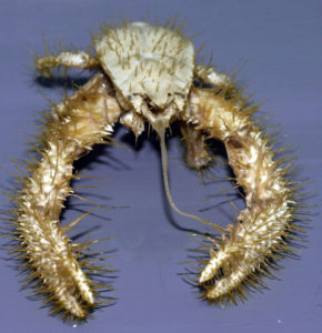
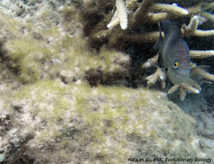
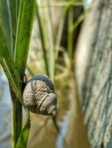

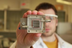

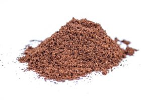
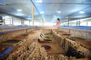


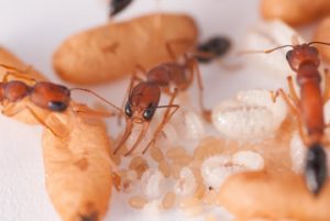
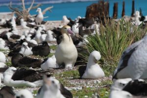
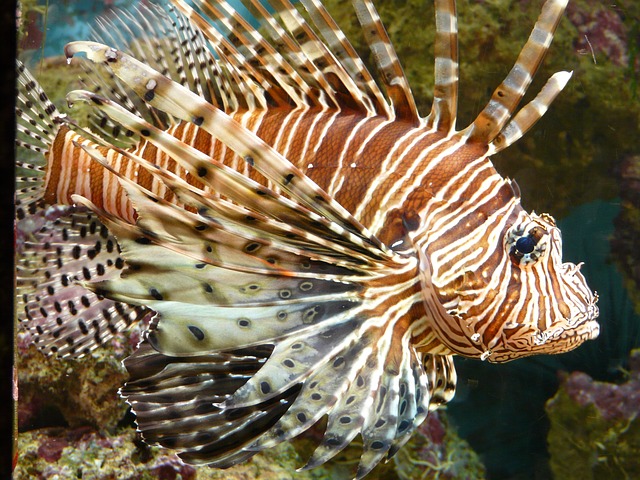

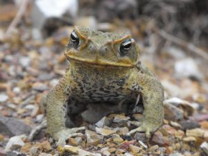
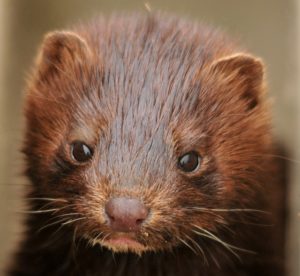
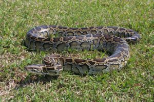


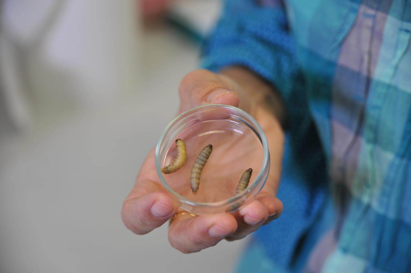

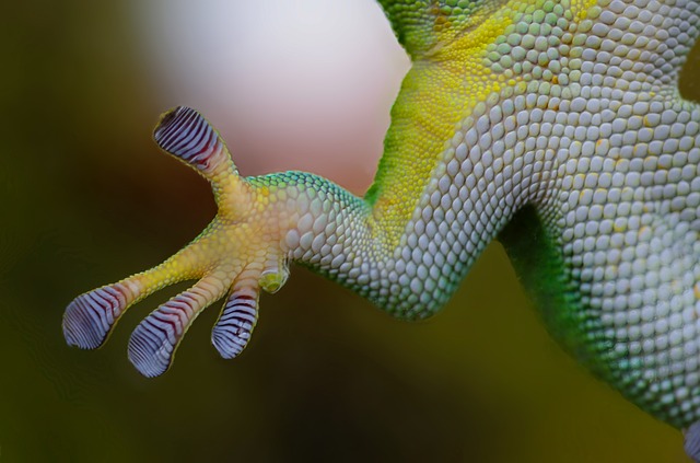
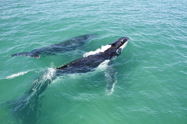


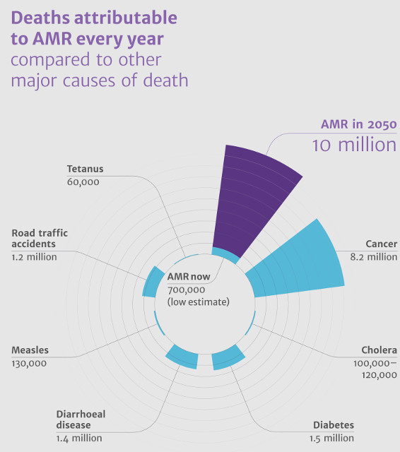
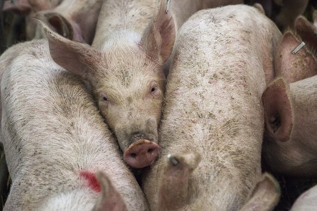

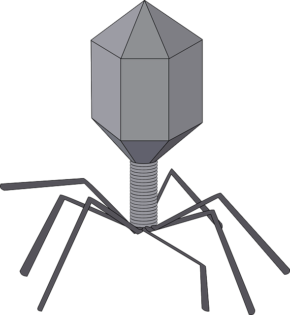

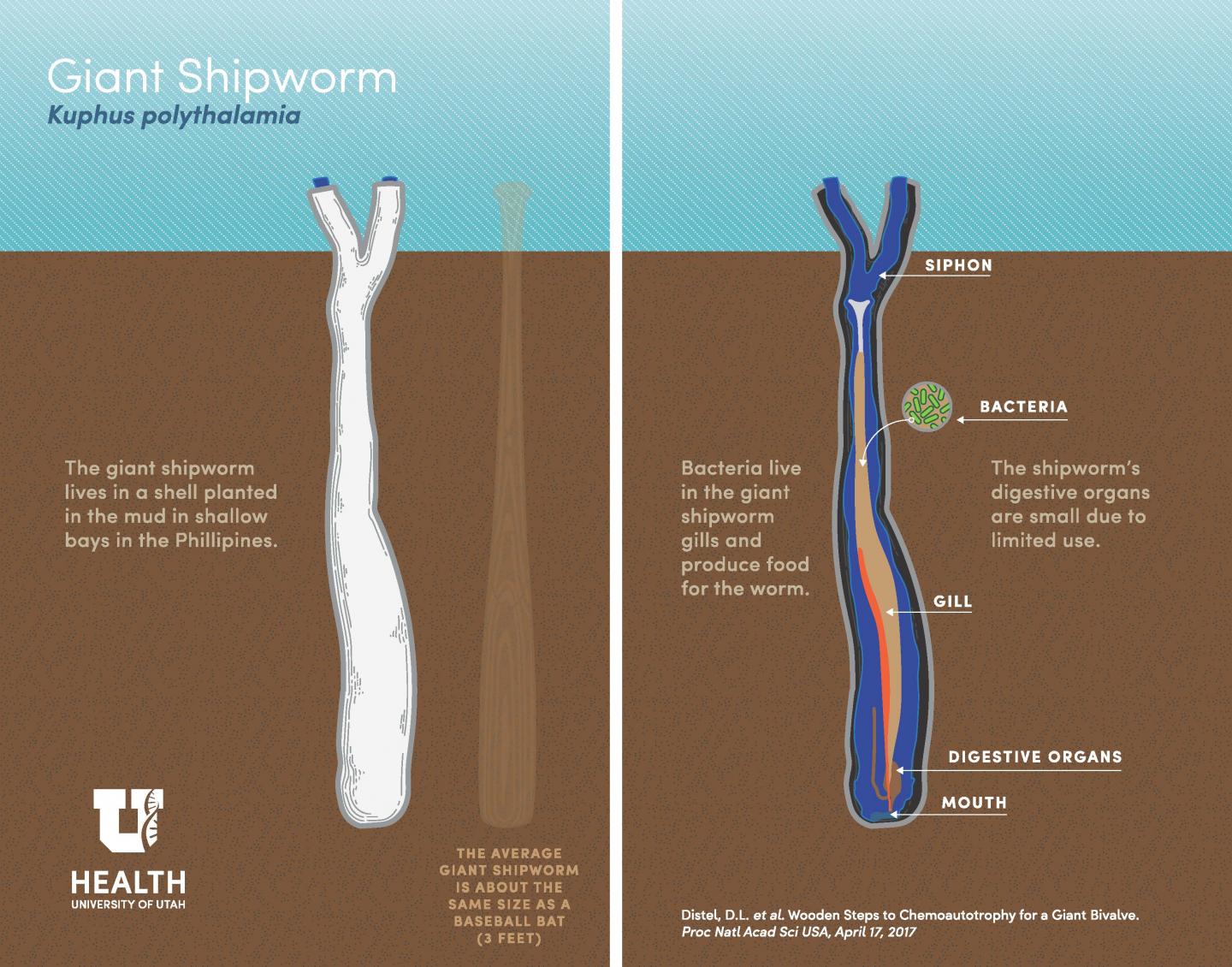
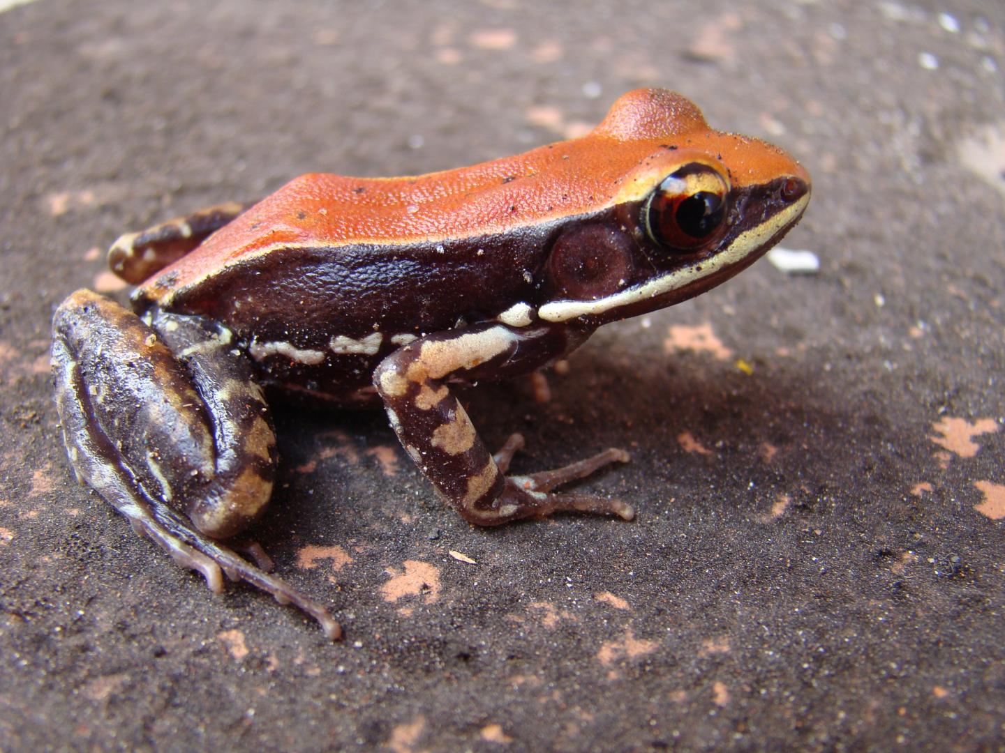
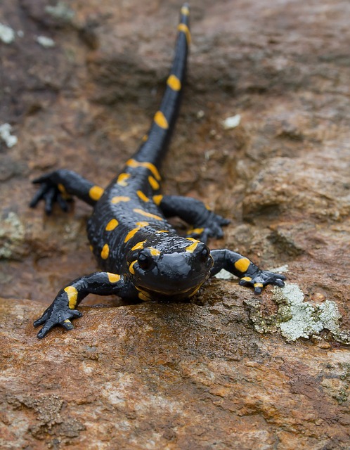

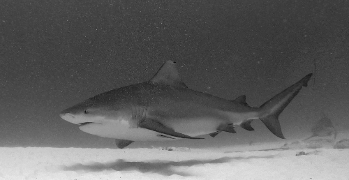
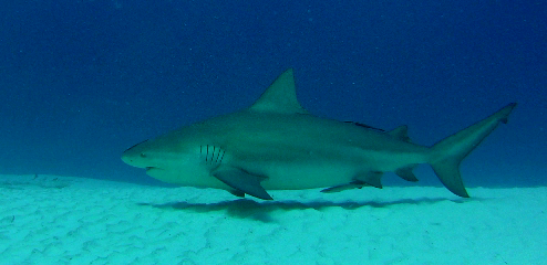
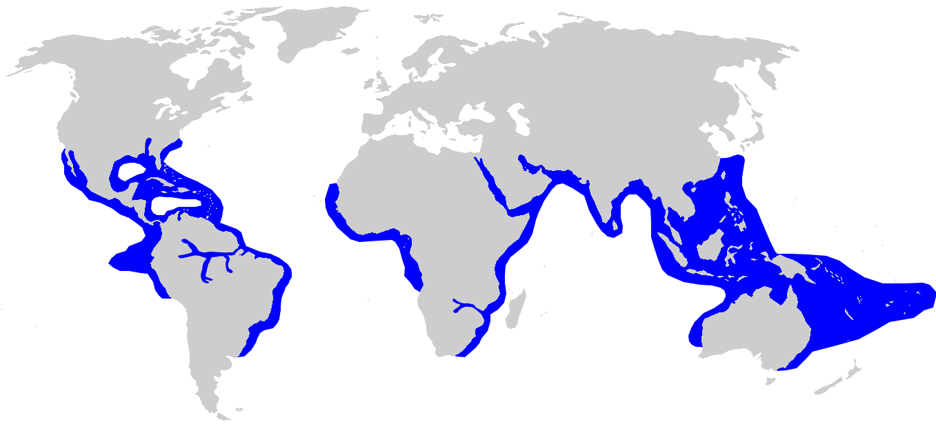
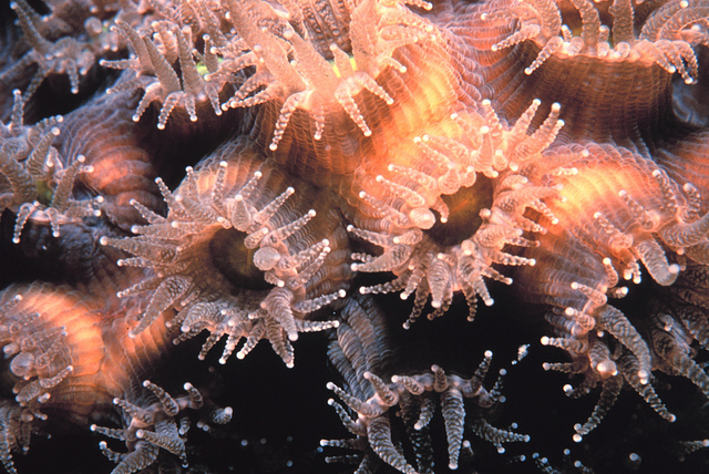
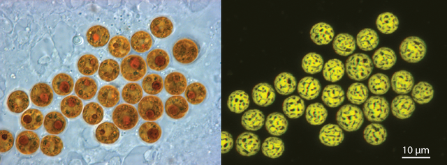
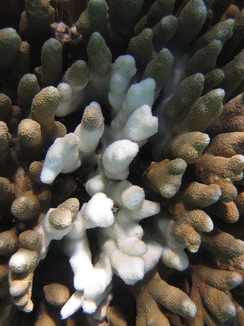
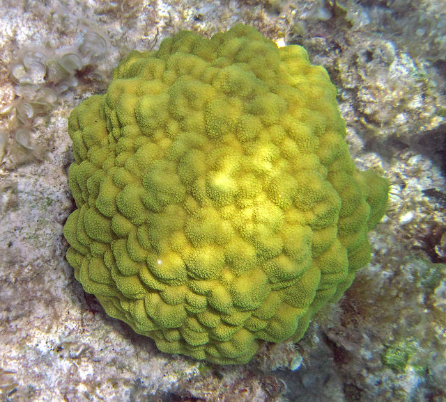

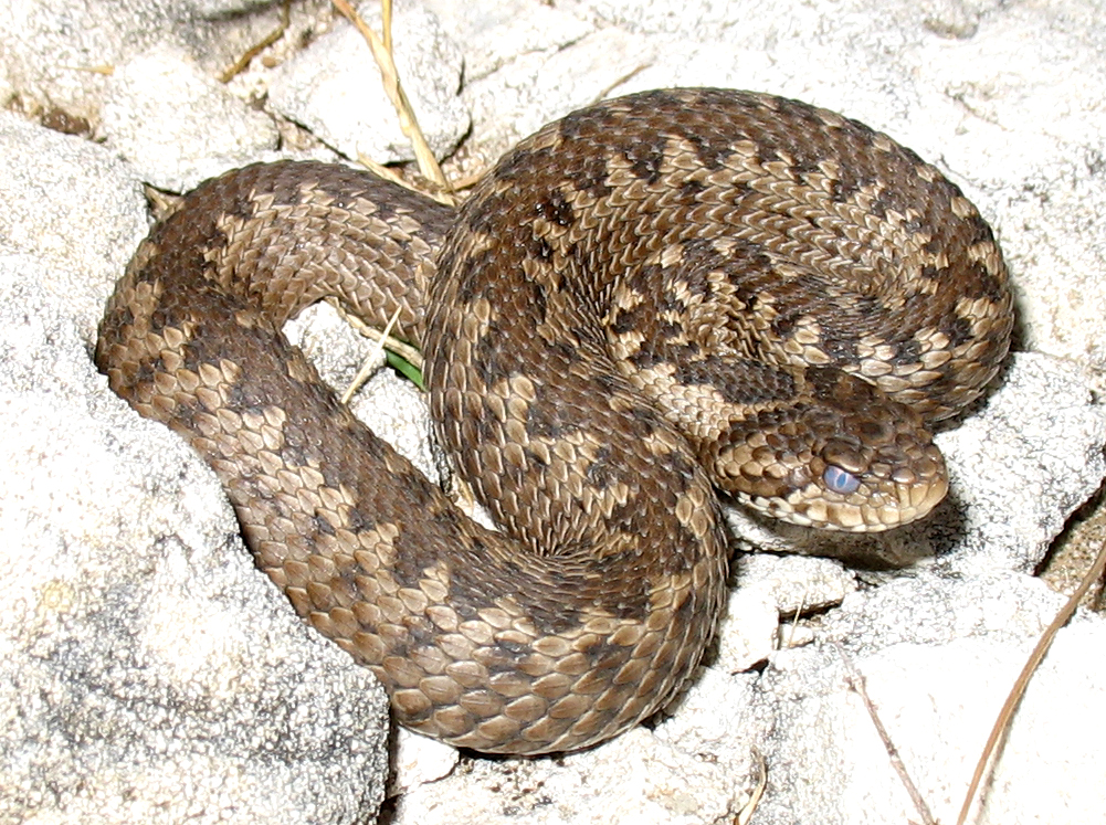

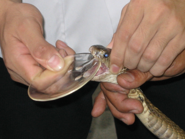